| Aim:
Application of magnetic nuclear spin resonance in medical diagnostics. |
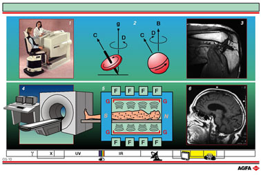
Diagram 1
shows a small Magnetic Resonance apparatus with which only the limbs of
the body can be investigated.
It consists of a sitting / lying table, a magnet tunnel and an imaging
and control console. It is normally equipped with a permanent magnet of
0.3 tesla.
Diagram 2 illustrates the similarity in properties between a charged atomic
nucleus (right) and a gyroscope (left).
A spinning top (gyroscope) spins around its axis and has an angular moment
C. In the presence of a gravitational field, (g), the gyroscope exhibits
a precession movement (D).
Some charged atomic nuclei, e.g. 1H, have magnetic properties that can
also be described by spin and magnetic moment (C). In the presence of
an external magnetic field (B) the individual spins of the H atoms present
can only take up certain orientations, in accordance with their energy.
Here too the spins exhibit a precession movement round the magnetic field.
At equilibrium a magnetic moment M that is lined up with the axis of the
external magnetic field will result for a large population of H atoms.
If an energy pulse (excitation) is supplied via a second magnetic field,
which is perpendicular to the first (radio frequency transmitter) then
the equilibrium is broken: the M vector changes in both size and orientation.
Following the disturbance caused by such a pulse the population of H atoms
returns to equilibrium. This equilibrium recovery can be tracked by measuring
the total magnetic moment as a function of time (relaxation); the relaxation
behaviour and the relaxation time give information about the magnetic
characteristics of the H atoms and their surroundings.
Diagram
3: MR-image of a bent knee that was reconstituted on either a monitor
and/or on film via a laser printer.
This has been achieved using the recorded relaxation times that are then
converted using suitable algorithms.
Relaxation measurements give an insight into the tissue structure and
the distribution of the components in an organ, head or limb.
Diagram 4:
shows a multi-functional classic machine in which the gantry ring (tunnel)
is completely closed. Just as with the limb-MRI scanner, MRIs now exist
with a broken circle to make investigations easier for claustrophobic
patients. These mostly use super-conducting magnets of up to 1.5 Tesla.
Just as in the CT the workstation / console is equipped with a powerful
computer, a mobile table and a gantry with a magnet, radio frequency pulser
and receiver.
In order to reduce interference and to protect the surroundings from the
high magnetic field a Faraday cage surrounds the scanner.
Diagram 5: a cross-section of the tunnel with a patient in it. Under the
influence of a strong magnetic field, the H atoms (the most prevalent
atoms in the human body) are forced to orientate along one directional
axis. When this magnetic field is interrupted relaxation occurs, and is
detected by the radio-frequency pulser/receiver and transmitted in digital
form to the computer. The computer processes the information further to
produce an analogue image.
Diagram 6:
MRI image of a skull.
Unlike radiography- or CT-investigation this method does not use X-rays.
Moreover, CT and MR provide complementary image-information. Classic radiology
still produces a projection of a surface structure onto a background structure.
Finally, only MR is able to give both an anatomical, as well as functional
and physiological, status.